
The dense connective tissue is divided into dense regular connective tissue and dense irregular connective tissue based on fibers arrangement. You will find the collagen fibers arranged in a definite plan in the histology of dense regular connective tissue.
Hi, if you want to know the histology of connective tissue in details, you might also know the histological features of dense connective tissue. This short article will discuss the histology of dense regular connective tissue with their location and functions.
You will also know the important identification points of dense regular connective tissue under the light microscope. If you are interested in learning the basic of dense connective tissue histology under the light microscope, continue this article.
Dense connective tissue histology
I hope you know the histological features of common connective tissue (like loose connective tissue). In any connective tissue, there are connective tissue cells, connective tissue fibers and amorphous ground substances. In dense regular connective tissue, you will find more densely packed and thicker collagen fibers along with less connective tissue cells and ground substances.
If you wish to identify the dense regular tissue slide under the light microscope, you might find out the following histological features from that slide.
#1. Compact bundles of collagen fibers
#2. Rows of fibroblasts
#3. Fewer ground substances in the tissue matrix
#4. Nuclei of fibroblasts
#5. Interfascicular connective tissue (if present in the tissue sample section)
#6. Blood vessels on the sample tissue section
Great, now you might try to identify the main histological features from the sample dense regular tissue section.
Identification points of dense regular tissue
You may ask to identify the dense regular connective tissue under the light microscope. The following identification points will help you understand and identify the dense regular connective tissue under the light microscope.
#1. The sample tissue section shows the compact, parallel bundles of fibers (especially the collagen bundles).
#2. Presence of parallel rows of fibroblasts in between the compact and thicker collagen bundles of the sample tissue.
#3. Less amount of ground substances are found in the sample tissue due to more compact and thicker collagen bundles.
So, this is a slide of dense regular connective tissue. You might add other possible identifying characteristics if you need them.
Histological features of dense regular tissue
If you think you should know the histology of dense regular connective tissue, you may continue this article. Here, I will discuss the cells (especially fibroblasts) and dense regular connective tissue fibres with their location and functions.
Fibers structure in dense regular tissue
This type of dense tissue contains the thicker and more densely packed fibers, especially the collagen bundles arranged in a definite plan. These fibers have distinct fibrous structures that show a regular and parallel arrangement.
Again, these collagen fibers have shining white appearances within the matrix. They show longitudinal striation, and if you perform the cross-section, you will find a finely dot area in the section.
Fibroblasts in dense regular connective tissue
You will find the less numbers of cells and ground substances in the matrix of dense regular connective tissue. Because of the dense arrangement of the collagen fibers or bundles, you will find the little amount of ground substances.
Between the thicker and densely packed collagen bundles, parallel rows of fibroblasts are found. These fibroblasts are the principal cells in dense regular connective tissue. The cytoplasm of these fibroblasts stained darkly with the basic dye.
If you don’t know about the histological features of fibroblast (most predominant connective tissue cells), you may read the details guide from anatomy learner. For your kind information, I would like to enlist the important histological features of fibroblast –
#1. Fibroblasts are the dominant cells of connective tissue that are flat and fusiform shaped with slender cytoplasmic processes.
#2. It contains the oval nuclei with the prominent nucleolus
#3. Often associated with the bundles of collagen fiber of the connective tissue
#4. They are two types – inactive fibrocytes and active fibroblasts
#5. Responsible for the formation of fibers of connective tissue and ground substances.
You may also find the interfascicular connective tissue in the dense regular tissue containing the fibroblasts and blood vessels.
Location, examples of dense regular connective tissue
This types of connective tissue usually found in tendons and ligaments structures. You may also find this dense regular tissue in fascia, aponeuroses and cornea structures.
#1. In tendons and ligaments structure
#2. In fascia
#3. Aponeurosis and cornea structures
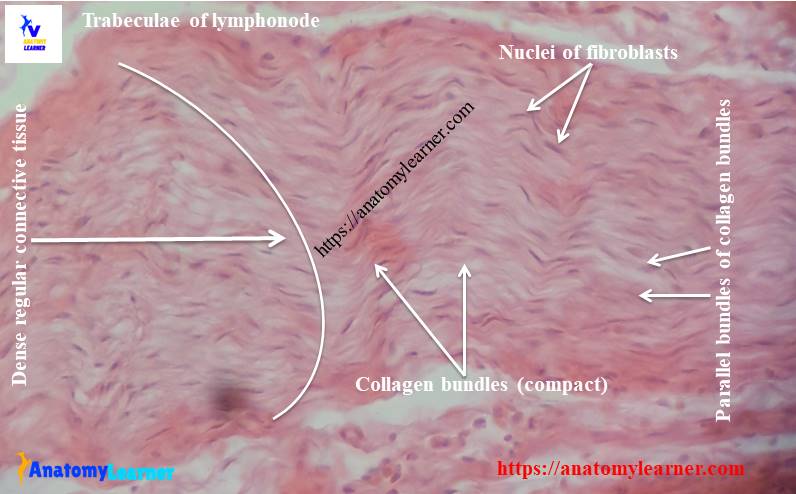
Slide pictures and diagram of dense regular tissue
Here, I shared the slide pictures and labeled diagram of dense regular connective tissue where I showed the thick and compact bundles of collagen fibers and parallel rows of fibroblasts.
If you want more slide pictures or diagrams of dense regular tissue, follow anatomy learners on social media (for more pictures of dense regular tissue). If you found any mistakes on the labeled diagram or real slide pictures of dense tissue, please let me know. I highly appreciate your support and will try my best to modify sample pictures.
Dense irregular connective tissue
You might also know the histological features of dense irregular connective tissue in details for further study on connective tissue. If you think you will also know the basis of dense irregular connective tissues with anatomy learner, you may read the details guide.
Loose connective tissue structure
If you are a beginner in connective tissue learning, you might know the histological features of loose connective tissue in details. I have published an article on loose connective tissue histology with real slide pictures and labeled diagram here in anatomy learner. You may know the structures, cells and fibers histology from loose connective tissue.
Elastic connective tissue
You know, elastic tissue presents in the few parts of an animal’s body that are yellow (microscopically). You will find the thick, refrigerant, rounded or flatten coarse parallel fibers in elastic connective tissue histology. Want to know more about elastic tissue histology?
You might also read other different articles related to connective tissue or other different organs system histology with anatomy learner.
#1. Cells of connective tissue with diagram
#2. Different types of epithelia with slide pictures under the light microscope.
#3. Histological characteristics of general tubular organs or hollow organs of animals
Conclusion
This guide will help you know the basic histology of dense regular connective tissue with real slide pictures and a labeled diagram. The identification points might help you to identify the dense regular tissue under a light microscope.
If you think this article is helpful and learn from this article, you may share it with your friends who also wish to learn basic connective tissue histology. Thanks for staying with anatomy learner and learning histology with me.