Cow Hoof Anatomy – Corium, Wall, Sole, and Bulb

Cow hoof anatomy labeled diagram

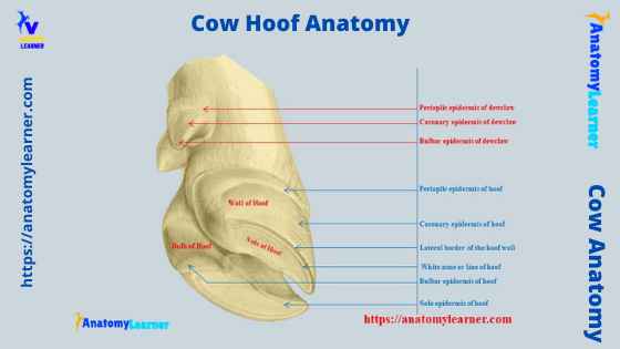
The cow hoof anatomy is a cornified modification of the epidermis that lies under the vascular layer and covers the end of the digits. You will find two primary and two accessory hooves on each limb of a cow. The leading hoof of a cow comprises three parts – periople, wall, and sole. Here, I will show … Read more

Pseudostratified Columnar Epithelium under a Microscope with a Labeled Diagram

Pseudostratified columnar epithelium

The pseudostratified columnar epithelium comprises a single layer of cells but seems to be multilayered. It is because different cellular heights and nuclei are also placed at a different levels. I will show you the pseudostratified columnar epithelium under a light microscope with its identifying points and labeled diagram.  I will also tell you the location of pseudostratified columnar … Read more

Simple Squamous Epithelium under a Microscope with a Labeled Diagram

Simple squamous epithelium under microscope labeled

Simple squamous epithelium under a microscope consists of a single layer of thin, flat, and scale-like cells. These cells are joined together by an intercellular junction and rest on the basement membrane, whose thickness depends on the location. Here, I will show you what simple squamous looks like under a light microscope.  Again, I will also tell you almost … Read more

Neuron under Microscope with Labeled Diagram

Neuron under microscope labeled

The structural and functional unit of the nervous system is the neuron that may easily observe under a light microscope. Neurons may vary considerably in size, shape, and other features. Here, I will provide details information and identifying points so that you may easily identify the neuron under a microscope.  A neuron consists of a cell body that gives … Read more

Dog Nose Anatomy – External Nasal Planum and Turbinates with Diagram

Dog nose anatomy (nasal cavity)

The dog nose anatomy comprises external features and the structures of the nasal cavity. From the external part of a dog’s nose, I will show you the features of cartilages, vomeronasal organs, and ligaments. Again, you will find the details anatomy of the nasal conchae, meatus, paranasal sinuses, and glands from the dog’s nasal cavity.  Throughout this … Read more

Cat Ear Anatomy – External, Middle, and Inner Ears with Diagram

Cat ear anatomy (diagram)

The cat ear anatomy consists of external, middle, and inner regions. You will find a pinna, external acoustic meatus or ear canal, and Henry’s pocket in the outer region of the cat’s ear. Again, the middle region of the cat’s ear consists of the tympanic membrane, cavity, and auditory ossicles.  The inner ear is located within the … Read more