Prophase Under Microscope – from Mitosis and Meiosis Stages
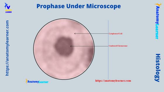
The prophase under a microscope shows the gradually becoming condensed chromatin, resulting in the formation of the individual chromosome. You know this prophase is the first stage of mitosis cell division which may quickly identify with the help of a light microscope. Again, you will also see the prophase 1 and prophase 2 stages in … Read more