Epididymis Histology Slide and Identification Points with Labeled Diagram

Epididymis histology

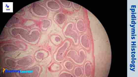
The epididymis is the comma-shaped structure in the male organ system that divides into head, body, and tail. It is made of highly coiled, tortuous ductus tubules and vascular connective tissue. In epididymis histology, you will find pseudostratified columnar epithelium lining and a circularly arranged smooth muscle layer beneath that epithelium.  Here, I will show you all … Read more

Ileum Histology Slide with Labeled Diagram and Identification Points

Ileum histology slide diagram

The ileum histology slide consists of the four layers like tunica mucosa, submucosa, muscular, and serosa. Here, I will show you the detailed histological features of the wall of the ileum slide with a labeled diagram. I will also provide you with the important identification points for the ileum histology slide so that you may identify it so quickly.  … Read more

Jejunum Histology Slide with Labeled Diagram and Identification Points

Jejunum histology slide labeled diagram

The jejunum is the second part of the small intestine of an animal. In the jejunum histology, you will find the four different layers like a tubular organ with some other specific identifying features. Here, I will show you the different features of a jejunum histology slide with a labeled diagram.  I will also describe the jejunum anatomy … Read more

Rumen Histology Slide Identification with Labeled Diagram

Rumen histology slide identification

The stomach of ruminants comprises four structurally distinct parts. Histologically, you will find a non-glandular mucosa having keratinized stratified squamous epithelium in the first three parts (rumen, reticulum, and omasum) of the ruminant stomach. I will show you everything about the rumen histology slide with their identification point and labeled diagram.  I will also show you the … Read more

Duodenum Histology Slide with Labeled Diagram

Duodenum histology diagram

There are three different parts –duodenum, jejunum, and ileum in the small intestine of an animal. You will find some salient microscopic features of each region of the small intestine. In this short article, I will discuss duodenum histology in detail, along with its slide identification points.  I will also show you the fundamental difference in the … Read more

Artery Histology – Elastic and Muscular Arteries Slides

Artery histology diagram

The blood vessels of the macrovasculature include arteries and veins. Arteries of the macrovasculature carry the blood from the heart to the microvasculature. These arteries are commonly defined by their position and further characterized histologically by their structure. In this short article, I will discuss artery histology with a labeled diagram.  First, I will describe the general … Read more