Skin Histology Slide Identification – Thick and Thin Skin Microscope Slides and Labeled Diagrams

Skin histology slide

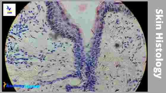
Skin is the heaviest organ of an animal’s body that covers the surface of the body. It comes into direct contact with the external environment. In the skin histology slide, you will find two main layers – epidermis and dermis. If you want to identify and understand the histological features from both thick and thin skin histology … Read more

Histology Slides Identification from Different Organ Systems

Histology slides identification

As a human medical student or veterinary student, you might learn histology slides with proper identifying characteristics. This article will show you the essential histology slides from the different organ systems of an animal’s body.  Here, you will get the histology slides of epithelial tissue, digestive system organs, respiratory organs, endocrine glands, male and female genital organs, and … Read more

Endochondral Ossification Process with Examples and Diagrams

Intracartilaginous ossification

In endochondral ossification, bone is formed by replacing the calcified cartilage. In this article, I will discuss the detailed process of endochondral ossification with labeled diagrams.  Hi there, welcome back again, and many, many thanks for getting into this article. I hope this article is going to be one of the best and easiest articles on the internet … Read more

Intramembranous Ossification Process with Diagrams

Process of ossification

Intramembranous ossification is the process of bone formation directly from mesenchyme. In this guide, I will discuss the steps or process of intramembranous ossification with a labeled diagram.  If you are interested in learning the process of intramembranous ossification in detail, you may continue this guide.  You will also know about the term – ossification center, osteoid, and mineralization here in this guide. After completing … Read more

Cardiac Muscle Histology and Slide Identification Points

cardiac muscle cover image

Cardiac muscle shows many structural and functional characteristics intermediate between skeletal and smooth muscles. Today, in this short article, I will show you the important histological features from the cardiac muscle histology slide.  You will get the basic guide to learn cardiac muscle histology with real slide images and labeled diagrams. I will also enlist the functions and identification … Read more

Skeletal Muscle Histology Slide Identification and Labeled Diagram

Skeletal muscle slide

The skeletal muscle fibers are elongated, cylindrical and multinucleated cells whose length may vary in different animals. In this short guide, you will get a basic concept of skeletal muscle histology from the real slide and labeled diagram.   You will also get the identification points of skeletal muscle histology slide with a little description here in this guide. I … Read more