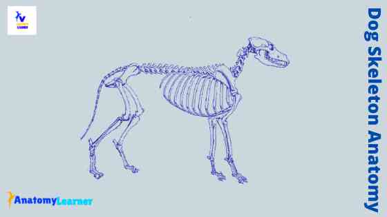
Dog Mouth Anatomy – Lip, Cheek, Oral cavity, and Salivary Glands with Diagram
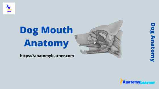
The dog mouth anatomy includes the lip, oral cavity, and associated structures. But, the term mouth includes only the opening between the lips into the vestibule of the oral cavity. Here, I will describe the anatomy of the dog’s mouth, including the lips and different parts of the oral cavity, with a labeled diagram. So, after reading this … Read more