Dog Neck Anatomy – Bones, Muscle, Glands, Veins, and Other Organs with Labeled Diagram

Dog neck anatomy diagram

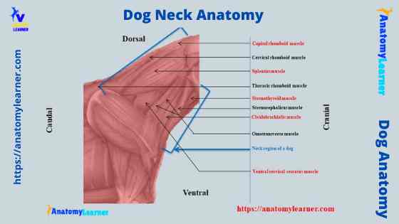
The dog neck anatomy consists of bones, muscles, glands, blood vessels, lymph nodes, and other essential organs. It is very common to find different injuries in the dog’s neck bones, muscles, and subcutaneous tissue. Again, you may find severe obstruction in the neck (in the esophagus or trachea) of a dog.  As a veterinarian, you might handle … Read more

Ruminant Reticulum Histology Slide with Labeled Diagram

Ruminant reticulum histology slide

The reticulum is a non-glandular part of the ruminant stomach. In the ruminant reticulum histology, you will find some permanent interconnecting mucosal folds known as reticular crests. I will show you every essential histological feature from the ruminant reticulum histology slide with labeled images.  I will also show you some differences among the three different parts of the ruminant … Read more

Cecum Histology Slide with Labeled Image and Diagram

Cecum histology slide labeled

The cecum histology slide follows the general pattern of the small intestine with some exceptional features. You may find a variation in the size of the gross cecum among the different species. But, in all domestic mammals, the cecum histology slide shows a substantial number of lymphatic nodules scattered throughout its length. Here, I will show you all the … Read more

Cow Skull Anatomy – Osteological Features of Cranial and Facial Bones of Ruminant Skulls

Cow skull anatomy labeled

The cow skull anatomy comprises cranial and facial bones. Most of these bones are irregular in shape and united immovably with the help of sutures (except the mandible). You will find some paired bones and single bones in the cow skull anatomy.  The cranial bones of the cow or ox skull from the cranial cavity accommodate the brain, different … Read more

Parathyroid Gland Histology with Microscope Slide Image and Labeled Diagram

Parathyroid gland histology slide

There are two or more pairs of parathyroid glands located on the posterior surface of the thyroid gland. You will find two main types of cells (chief and oxyphils) in the parathyroid gland histology slide. Here in this article, I will show you all the structures of a parathyroid gland with a microscope slide image and a … Read more

Tonsil Histology Slide with Labeled Diagram – Histological Features of Palatine and Lingual tonsils

Tonsil histology slide microscope image

Near the junction of the oral cavity and the pharynx, you will find several collections of lymphoid tissue that refer to the tonsil. In tonsil histology, different essential structures like tonsillar crypts, lining epithelium, numerous lymphatic nodules, and dense connective tissue capsules should be identified under a light microscope.  I will show you all these structures from … Read more